![]()
![]()
 Note: Be sure to review the history of the microscope and microscope safety procedures. Links are provided at bottom of page. For a printer friendly version, click here.
1.
Ocular Lens (Eyepiece) - where you look through to see the
image of your specimen. Magnifies
the specimen 10X actual
size.
2. Body tube - the long tube that supports the eyepiece and connects it to the objectives.
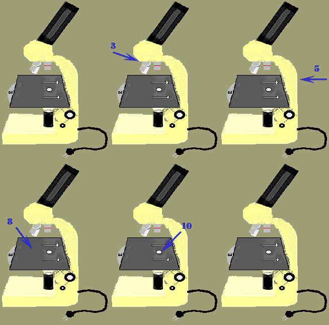
3. Nosepiece - the rotating part of the microscope at the bottom of the body tube; it holds the objectives.
4. Objective Lenses - (low, medium, high). Depending on the microscope, you may have 2, 3 or more objectives attached to the nosepiece; they vary in length (the shortest is the lowest power or magnification; the longest is the highest power or magnification).
5. Arm - part of the microscope that you carry the microscope with; connects the head and base of the microscope.
6. Coarse Adjustment Knob - large, round knob on the side of the microscope used for "rough" focusing of the specimen; it may move either the stage or the upper part of the microscope. Location may vary depending on microscope - it may be on the bottom of the arm or on the top.
7. Fine Adjustment Knob - small, round knob on the side of the microscope used to fine-tune the focus of your specimen after
using the coarse adjustment knob. As
with the Coarse Adjustment Knob, location may vary depending on the
microscope. placed on the stage for viewing.
9. Stage Clips - clips on top of the stage which hold the slide in place.
10. Aperture - the hole in the stage that concentrates light through the specimen for better viewing.
11. Diaphragm - controls the amount of light going through the aperture; may be adjusted.
12. Light or Mirror - source of light usually found near the base of the microscope; used to direct light upward through the microscope. The light source makes the specimen easier to see.
< Bacteria & Plants > < Cells & Heredity > < Human Biology & Health > < Microscope History > < Microscope Lab > < Microscope Safety > < Website Directory > |